

ABOUT US
Time:2020-11-14 Views:300
Introduction to the main points of the fluorescent Leica stereo microscope
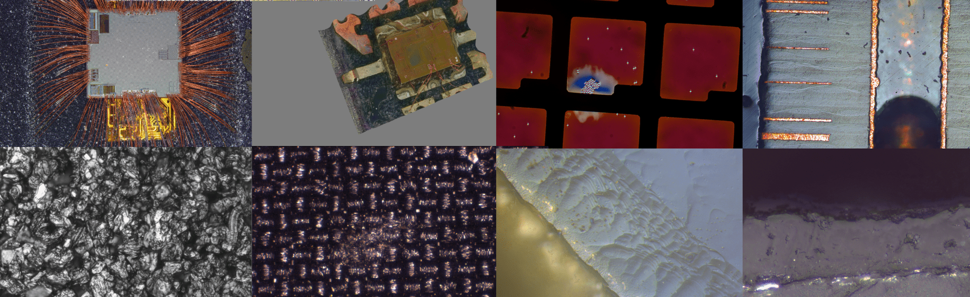
The fluorescent Leica stereo microscope is a major development in general microscopy. It has unique advantages. Fluorescence three-dimensional microscopy is to observe the various images of the standard bars under the background of the black reading. It has a good anti-bow, so the observation is clear and convenient. Brother Yi: identification, the eyes will not be tired. Because the concentration of fluorescent chromatin is extremely dilute (Meter 1: Iooo00), it can emit bright fluorescence. Therefore, the general oil body is said to be motionless. It is for observation in vivo, such as studying the process of functional organs and subtle structures, and certain metabolic processes. The changes, etc., provide a wide range of possibilities. In addition, the Yingguang dyeing method is relatively simple to make time and road signs, which is especially suitable for rapid work such as rapid diagnosis of infectious diseases.
| ") | ") |
| test | ||
| test | ||
| test | ||
| test | ||
| test | ||
| test | ||
| test |
The fluorescence microscopy of the fluorescence stereo microscope can be used to change the bright field to the dark field, but it is generally better to use the right field, because the scene background it produces is black, the image contrast is strong, and there is no bright field. Convenience. As for transmitted fluorescence or reflected fluorescence, in general, reflected fluorescence has a wide range of applications because it is not only suitable for observing ordinary markings, but also for observing unclear specimens.
Fluorescence Leica stereo microscopes can be excited by ultraviolet light or violet blue light according to the characteristics of excitation light wavelength. The feature of the outside light is to observe the autofluorescence of body tissues <such as nucleic acid and diamond, etc.). It is convenient to distinguish the background fluorescence from the fluorescence of the staining target in the organization d:. But the shame is: often cause mark 4; fluorescence photoquenching, the specimen is difficult to observe repeatedly, and the sand photomicrography is difficult to group. In addition, it has a significant killing effect on living specimens, and it also hurts the eyes of people who have been engaged in this work for a long time. Especially for most fluorescent pigments, the ultraviolet region is not the maximum spectral absorption wind and therefore cannot be excited first. The purple blue light does not have the above-mentioned shortcomings, but the shortcoming is that it cannot stimulate the autofluorescence of the tissue, so it is not suitable for studying the white body fluorescence structure of the tissue.
Fluorescence stereo microscope for individual fluorescent pigments, known as Rhodamin R200, can use longer wavelength blue and green light as excitation light.
Results The fluorescence images observed by the fluorescence Leica stereo microscope were mainly based on two indicators: Liu's results, one was the morphological characteristics, and the other was the brightness of the fluorescence. In the immune fluorescent work towel, this is especially the case. The two must be combined to make a comprehensive judgment to not be partial. Correction records are based on subjective indicators or objective indicators, but they are usually subjective indicators observed by the worker's daily effort. Because this is a qualitative observation, the accuracy of the record is poor. With the development of science schools, the use of objective indicators to record results has begun to be applied.
1. Use light-sensitive battery leakage meter Z; the brightness of the image, or be combined with a scanning device to scan a specific part of the field of view, such as a fluorescent metallographic photometer;
2. Insert neutral filters of different order into the light path between the excitation filter and the specimen to see the extremes of the fluorescence reduction;
3. Directly use a fluorometer to measure the fluorochrome and the fluorescent stained specimen under the microscope;
4. Different specimens are photographed with the same meteorological time, and then the film image is measured with a densitometer.


Links:

Inquire Now
Inquire Now
×Hello,please leave your information below,we will contact you asap.


")


